Novel Remyelination Assay Allows High-Throughput Drug Screening
A new technology uses the actual myelin-making behavior of cells to look for drugs that may promote remyelination and prevent disability-causing nerve damage in people with MS. The first candidate is in clinical trials.
In the brains and spinal cords of people with multiple sclerosis (MS), the repair of demyelinating lesions remains inexplicably complex. But a new screening platform makes it look easy. Stripped down to the basics, an individual oligodendrocyte will wrap myelin around anything that feels like an axon, including tiny plastic fibers and microscopic glass pillars.
This single-minded drive of myelin-making cells forms the heart of a new high-throughput screening platform. Using the new platform, researchers found several drugs already on the market for other conditions that boosted the cells’ myelin production (Mei et al., 2014). One candidate, clemastine, an over-the-counter antihistamine, is now in a phase 2 clinical trial in people with relapsing-remitting MS (RRMS).
The findings were published online July 6 in Nature Medicine by researchers in the lab of Jonah Chan, Ph.D., a neuroscientist at the University of California, San Francisco (UCSF), and his collaborators.
“A lot of people are trying to find ways of identifying remyelination therapies, and this is a unique approach,” said neuroscientist Wendy Macklin, Ph.D., professor and chair of cell and developmental biology at the University of Colorado School of Medicine, in an interview with MSDF. “What Jonah has is something that really does look like it’s behaving like a myelinating cell.”
![This extreme close-up with a scanning electron microscope shows oligodendrocytes cultured in a well among the tapered glass micropillars. Reprinted by permission from Macmillan Publishers Ltd.: <em>Nat. Med.</em>, 2014 July 6 (doi 10.1038/nm.3618, copyright 2014)].](http://www.msdiscovery.org/sites/default/files/1g_resized.jpg) Of the first 1000 compounds screened by Chan’s team, the most potent were a cluster of eight FDA-approved drugs that bind to muscarinic receptors, better known for their presence on neurons that regulate internal organs, such as the heart and digestive tract. A similar cluster, including benztropine, an obsolete Parkinson’s drug, popped up in a screen of more than 100,000 drugs reported last year (Deshmukh et al., 2013).
Of the first 1000 compounds screened by Chan’s team, the most potent were a cluster of eight FDA-approved drugs that bind to muscarinic receptors, better known for their presence on neurons that regulate internal organs, such as the heart and digestive tract. A similar cluster, including benztropine, an obsolete Parkinson’s drug, popped up in a screen of more than 100,000 drugs reported last year (Deshmukh et al., 2013).
“It’s always nice in science when two different approaches converge on the same observation,” said Luke Lairson, Ph.D., a biochemist at The Scripps Research Institute in San Diego, California, and senior author of last year’s report. Chan agreed: “It validates both sets of studies.”
The next generation of MS research
The high-throughput screening results come at a pivotal moment in MS research, when attention is turning to the major challenge of repairing myelin. Investigators hope that by repairing myelin they’ll also prevent the neuronal damage believed to underlie the progressive disability that eventually affects most people with MS.
“MS is an inflammatory and demyelinating disease,” said Bernard Zalc, M.D., Ph.D., a neuroscientist at the Hôpital de la Pitié-Salpêtrière in Paris, in March at the American Society for Neurochemistry meeting in Long Beach, California. “In the past 20 years, companies have made fantastic progress in the treatment of the inflammatory parts of the disease. Unfortunately, the repair of demyelinating lesions is awaiting therapeutic progress.”
Last week, Chan, Lairson, and Macklin spoke on new screening tools at a one-day remyelination conference at the New York Academy of Sciences. “We’re at a stage where we can start identifying small molecules in high-throughput screening with the potential to work,” Lairson said. “The next hurdle is the clinical evaluation of the agents.”
The standard screens for myelin-promoting compounds feature oligodendrocyte precursor cells. They test for agents that push them to develop, or differentiate, into myelin-making oligodendrocytes. The myelin is an extension of the membrane surrounding the cell. In the normal course of development, the oligodendrocyte pumps out extra membrane, which embraces neighboring axons and forms the insulating myelin sheath that speeds nerve conduction. The membrane is enriched with extra fatty lipids and different proteins, including myelin basic protein and others that the immune system attacks in MS. In the lab, researchers can label the cells to see a key genetic change at each stage.
By themselves in a lab dish, oligodendrocytes do not make myelin, but their membranes do show the initial genetic changes. “A lot of groups, including ours, have done the standard screens looking at the increase in the differentiation of primary cells,” Macklin said. “The unique element to Jonah’s screen is the added three-dimensional component, where he’s looking at the ability of cells to wrap myelin around little pillars.”
In their screening studies, both teams ran the leading candidates through additional tests—evaluating their myelin-making potential in oligodendrocytes cocultured with neurons and their ability to remyelinate axons in mouse models of demyelination. Macklin believes zebrafish can provide a rapid and inexpensive intermediate step between the culture dish and mice to evaluate the biology, timing, dosing, and toxicity of the candidate compounds, as well as molecular variations that could make them safer and more effective in people.
Taking it to the clinic
In the meantime, compounds with remyelination potential that are already approved by the U.S. Food and Drug Administration (FDA) provide a helpful shortcut in testing them in people, UCSF neurologist Ari Green, M.D., told MSDF. Green is leading a phase 2 clinical trial to test clemastine in 50 people with RRMS. The study started in January 2014 and is approaching 50% enrollment, with a goal of finishing in late 2014. The team has a strict selection criterion: people with significant myelin loss but with intact axons that can be remyelinated. The FDA’s granting of a New Drug Application exemption expedited the trial, allowing researchers to study a drug in a condition for which it was not originally approved, according to a UCSF news release.
“We can skip the phase 1 testing, because we know it’s safe overall in people on average,” said Green, a co-author of the new study. He cautioned, though, that clemastine may be dangerous in some subset of people. “We are using a higher dose than is used routinely in a background of people who have other symptoms,” he said. Drowsiness, the most common side effect of clemastine, likely rules out the approved molecule as a final drug, even if found safe and effective, Green told Xconomy, because fatigue is the number one issue for people with MS.
![A cluster of antimuscarinic compounds scored high in cell differentiation and membrane wrapping in the high-throughput BIMA screen. Reprinted by permission from Macmillan Publishers Ltd.: <em>Nat. Med.</em>, 2014 July 6 (doi 10.1038/nm.3618, copyright 2014)].](http://www.msdiscovery.org/sites/default/files/3a_resized.jpg)
Clemastine isn’t the only experimental aspect of the trial. It’s unclear how to measure remyelination. “We’re still trying to figure out what the right phase 2 clinical outcome is for an agent that is protective and restorative for myelin,” Green told MSDF. “Part of the trial design is to be somewhat agnostic. We have selected a principal outcome, but we’re open to the idea that it may not turn out to be the best and needs to be modified or adapted.”
The study’s primary outcome will be a test to measure the speed of a shining light from the retina to the visual cortex of the brain. Unmyelinated axons conduct electrical signals at about 1 meter per second, or the equivalent of 3 miles per hour, Green said. Myelin turbocharges signals along the axon up to 100 meters per second or 300 miles per hour. Secondary outcome measures include tolerability, brain MRI, and disability scores.
The study’s crossover design may yield additional clues about how often and how long to give the drug. Half of the patients will be on the drug for 3 months and then cross over to placebo for 2 months. The other half will start on placebo and cross over to clemastine.
“Some people ask, how do we know we will not overmyelinate?” Green said. “Those factors that serve as brakes on the system are still there. We’re not overcoming everything. We’re trying to drive things to a state where there is a more permissive environment for myelination.” A remyelinating agent may recapitulate one of the earlier developmental periods with a burst of myelin production, he said.
Making myelin on molehills
Chan credits his department chair and co-author Stephen Hauser, M.D., with the original vision for a high-throughput myelin-making assay. In fact, when Hauser first broached the subject while recruiting him 5 years ago, “I told him, ‘Impossible,’ ” Chan said. The standard myelinating culture using a rat neuron is too tedious and temperamental for fast and inexpensive mass screening, although Chan and Lairson employed the technique to confirm their top hits.
Chan turned to his basic studies of repair and remyelination, but the idea percolated in the back of his mind. “It’s such an unmet need in MS now,” Chan said. “There are great therapeutics for preventing the immune system from doing more damage, but there is nothing to promote remyelination.”
It used to be thought that axons sent some molecular signal to oligodendrocytes, instructing them to begin myelination. But one of Chan’s graduate students, Sheila Rosenberg, noticed that oligodendrocytes would form myelin even around dead axons (Rosenberg et al., 2008). At UCSF, he and his collaborators found that oligodendrocytes would also wrap myelin around thin plastic fibers (Lee et al., 2012).
“That step—nanofibers to model myelination—was a necessary prerequisite” to the new study, Chan told MSDF in an interview. “It told us the cells are capable of wrapping artificial structures. The nanofibers showed us that oligodendrocytes are sensitive to the biophysical properties of fiber diameter and shape.” But computer software could not reliably detect the newly formed myelin, making the fibers unsuitable for high-throughput screening. For the automated detection, the assay would also need a robust yes-or-no readout, he reasoned.
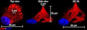
One night at home, Chan was using a new three-dimensional software program to play around with images of cells wrapping the nanofibers. As he turned the nanofiber this way and that, he caught a bottom-up view of the myelin around the shaft. “Here’s the trick,” he said. “If a cell has wrapped a single process around a pillar, it looks like a ring from the bottom. That’s when I realized that the myelin ring represents the quick way. Either you have a ring or you don’t.”
![Conceptually, Chan reasoned that the rings of fluorescent images taken from the bottom of the glass plate would allow for measurement of myelin between the base and tip of each pillar. Reprinted by permission from Macmillan Publishers Ltd.: <em>Nat. Med.</em>, 2014 July 6 (doi 10.1038/nm.3618, copyright 2014)].](http://www.msdiscovery.org/sites/default/files/1e_1f_combined.jpg)
Led by postdoctoral fellow Feng Mei, Ph.D., Chang’s team designed short micropillars that tapered at the top and lined them up on a glass plate like a platoon of squatty conehead soldiers. They started with a 96-well plate containing 10,000 micropillars in each well and now work with a 384-well plate.
 The micropillars are spaced with military precision far enough apart so that each oligodendrocyte can reach and wrap only one pillar. (In the brain and spinal cord, one oligodendrocyte typically reaches out to wrap short segments of about 15 to 20 nearby axons, although they have been known to embrace as many as 100 axons.) Imaged from the bottom of the slide, computer software can automatically detect a ring and quantify the results. Confocal microscopy captures five slices from the base to the tip.
The researchers call the technology BIMA, short for binary indicant for myelination using micropillar arrays. If that sounds vaguely spiritual, you are correct. The name references the Hebrew word for the raised platform on which the Torah rests. Chan chose the term to reflect the physical screening platform and the abstract idea of the next major step in MS translational research.
The proprietary technology has been licensed to a new drug-discovery company to follow up on the search for novel small-molecule remyelinating therapies for people with MS. The company, called Inception 5, is a partnership between the venture capital firm Versant Ventures and the biotechnology company Roche.
Back to the lab
Both the Chan and Lairson labs are looking at the mechanisms of action of the antimuscarinic candidates from their screens, as well as other ways they may be promoting myelination. Benztropine was the top performer in the Lairson team’s studies, but it has not progressed to clinical trials. The researchers are also evaluating newer antimuscarinic agents approved for other conditions. Lairson is pursuing two paths: “Can we find something that operates through antimuscarinic activity that’s safe, and are there alternative mechanisms that will work for which we can find novel compounds?” So far, the dual inhibition of the muscarinic receptors M1 and M3 appears to be required for differentiation and myelination, he said, and another unidentified target is likely involved.
Chan’s team has screened another 1000 compounds in the lab and identified and validated two additional clusters of potential remyelination agents. He also wants to use the micropillar assay to give him more basic information about oligodendrocytes and myelination, a poorly understood process in healthy mammal brains. BIMA “may provide insight into new pathways and receptors essential for oligodendroglia development and myelination,” the authors write in the paper.
“A lot of science depends on being in the right place at the right time,” Chan said. “I'm a basic neuroscientist surrounded by MS clinicians. The environment is set up so that we’re constantly thinking about ways to apply basic science to patients. It’s quite a collaborative effort to have the whole group here consider what is needed for patients with MS when they conduct research in the lab. Inevitably, we apply what we find in the lab to what is needed in the clinic.”
Key open questions
- Which specific pathways are the drugs identified by the screen acting on?
- How many different pathways exist that promote remyelination?
- Does a remyelination therapeutic need to hijack multiple pathways or just one of them?
- Once remyelination agents have been identified, when in the course of the disease will they be most effective, how long will they need to be given, and how will their effectiveness be measured?
Disclosures and sources of funding
Chan and his co-authors were supported by the U.S. National Multiple Sclerosis Society Harry Weaver Neuroscience Scholar Award, the UCSF CTSI Catalyst Award for Innovation, gifts from friends of the Multiple Sclerosis Research Group at UCSF, and the Joint Research Fund for Overseas Chinese Young Scholars. Chan and Green are co-founders and equity holders in Inception 5.
Macklin is funded by the U.S. National Institutes of Health and the National MS Society. She is on the scientific advisory board for Renovo Neural.
Lairson declares no competing interests.


Comments
A tremendous concept, a seminal article that probably will help in the ID of newer agents that will promote remyelination. However, we all recognize that significant axonal loss occurs even at the earliest stages of RRMS or CIS. With no or altered scaffolding for the oligodendrocytes to 'wrap around', this assay will hopefully trigger the use of appropriate agents that can stem further demyelination, early.
Use of medications early is critical in MS and this assay will help propagate that principle.
J Avasarala