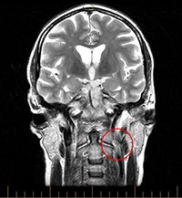
Abnormal Venous Outflow Dynamics May Not Be Implicated in MS
Imaging did not show chronic cerebrospinal venous insufficiency to be common or significant in MS patients
Altered venous outflow dynamics are not common or pathophysiologically significant in patients with multiple sclerosis (MS), according to an imaging study published online in advance of print in the Multiple Sclerosis Journal (Brod et al., 2013).
 “Chronic cerebrospinal venous insufficiency (CCSVI) was postulated as causally related to MS and disproportionately distributed among clinical MS disease phenotypes,” wrote Staley Brod, M.D., of the University of Texas Health Science Center in Houston, Texas, and colleagues. “Purportedly established by the presence of two or more disordered venous outflow parameters as measured by intra- and extracranial duplex ultrasound, CCSVI was originally reported as exclusively associated with the diagnosis of MS and was not found in other diseases or normal controls.”
Since 2010, results have been conflicting regarding the prevalence of CCSVI in MS and the occurrence of CCSVI in people without MS, due in part to use of varying imaging methods.
The present study sample included 206 patients with MS and 70 without MS enrolled in a single-center, prospective, case-control study. Investigators blinded to MS status compared neurosonography (NS), magnetic resonance venography (MRV), and transluminal venography (TLV) in subsets of these participants.
High-resolution B-mode NS included color and spectral Doppler of extracranial and intracranial venous drainage. Participants underwent MRV in a 3T scanner before and after injection of gadofosveset trisodium contrast.
Very Low CCSVI Prevalence
As determined earlier (Barreto et al., 2012), prevalence of CCSVI on NS was much lower than previously reported. Groups with or without MS had similar proportions of NS findings consistent with CCSVI (3.88% vs. 7.14%; p = 0.266).
Of 99 patients with MS who underwent systemic and intracranial MRV, 98 were evaluable, and 26 had discrepant MRV and NS findings. Four had abnormal NS but normal MRV.
Forty of 98 evaluable patients with MS also underwent TLV, including pressure measurements of the superior vena cava, internal jugular, brachiocephalic, and azygous veins. None had TLV pressure gradients suggesting clinically significant functional stenosis. However, 1 of 39 with accessible azygous veins had minimal narrowing, and 55% of internal jugular veins showed various degrees of stenosis.
“The three imaging approaches provided generally consistent data with discrepancies referable to inherent technique properties,” the investigators wrote. “Our findings lend no support for altered venous outflow dynamics as common among MS patients, nor do they likely contribute to the disease process.”
Mean age of MS patients was 47.6 years and mean duration of MS was 9.9 years. Nearly two-thirds (62) had relapsing-remitting MS; 23 had secondary progressive MS, five had primary progressive MS, six had clinically isolated syndrome, and two had progressive relapsing MS.
Comparison of Imaging Techniques
- NS allows noninvasive measurements in different postures and under different physiologic stresses but has limited access to select proximal and intrathoracic venous drainage system components.
- MRV, with or without contrast enhancement (CE), offers excellent access to the intracranial and proximal venous drainage system of the brain but is not ideal for time-of-flight visualization below the neck.
- TLV is considered the “gold standard” but is invasive and subject to flow artifacts due to catheter placement and vascular spasm.
“In our hands, both CE-MRV and TLV had advantages for accessing certain regions of the venous system that are poorly or not insonated by ultrasound such as poor temporal bone windows and the azygous system,” the investigators concluded. “Both CE-MRV and NS provided inherently different and complementary types of information, with generally congruent results. Dynamic CE-MRV may be preferable to TLV in screening for venous abnormalities because it evaluates the most proximal extent of the IJVs [internal jugular veins] and is less invasive.”
Key open questions
- What is the best protocol for venous drainage imaging among patients with MS?
- Are there subsets of patients with MS and CCSVI who differ clinically or in therapeutic response from those without CCSVI?
Image credit
Contributed by an anonymous patient under Creative Commons License.
Disclosures
The National Multiple Sclerosis Society (NMSS) supported this study. The investigators’ local Center for Clinical and Translational Sciences, funded by the National Center for Research Resources (NCRR), provided limited personnel support. None of the investigators disclosed any financial interests in the study outcome, but during the course of the study, various of the investigators received support for other, unrelated activities from Acorda, Astellas, Avanir, Bayer HealthCare, Biogen Idec, Celgene, the Clayton Foundation for Research, the Department of Defense, the Consortium of MS Clinics, Eli Lilly, EMD Serono, Genzyme, Hoffmann-La Roche, I-Tomography, Medcomp, Medscape CME, Millipore (Chemicon International) Corporation, National Institute of Drug Abuse, Novartis, Pfizer, Questcor, Sanofi-Aventis, Teva Neurosciences, and the Texas Neurological Society.


