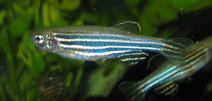
Zebrafish
Animal description
 The relatively simple zebrafish, Danio rerio, may not spring to mind as an animal useful for studying the complexities of multiple sclerosis. However, the animals are proving useful for studying the mechanisms involved in myelination and as a medium for high-throughput drug screening.
Currently, there is no multiple sclerosis zebrafish model per se. Researchers use the animals to study normal myelination processes to better understand them and to find potential targets for remyelinating therapies. But the immunopathogenic aspects of MS have not yet been characterized in zebrafish. According to David Lyons, Ph.D., of the University of Edinburgh in the U.K., this is partly because the adaptive immune system is still not well understood, though researchers have studied the innate immune system extensively.
Zebrafish make an appealing animal model because they are cheaper and easier to work with than mice. A zebrafish pair produces between 100 and 300 offspring, and the embryos develop outside the mother. Their axons begin to myelinate by day three, when they are still a tiny embryo, and they are sexually mature in about 8 to 12 weeks. Best of all, the embryos are completely transparent. This enables researchers to watch myelination take place in the central nervous system (CNS) in real time, with the help of fluorescent proteins. Because zebrafish share 70% of their genome with humans, the genetic and physiological processes of myelination are thought to be well conserved.
Transgenics
Relative to modifying the mouse genome, researchers can easily modify the zebrafish genome, and often at less expense. In MS research, scientists use transgenic zebrafish lines that express green fluorescent protein (GFP) in oligodendrocytes and oligodendrocyte precursor cells (OPCs). Using these, researchers have been able to visualize several aspects of the interaction between OPCs, oligodendrocytes, axons, and myelin in the developing zebrafish. Specifically, Kirby et al. (2006) used a transgenic line, Tg(nkx2.2a:megfp), to monitor OPC behavior in real time. Two other teams (Jung et al., 2010; Almeida et al., 2011) using similar transgenic methods used myelin basic protein in oligodendrocytes to drive GFP expression. Czopka et al. (2013) used a transgenic line to monitor myelination in real time. Most recently, Yin and Hu (2013) examined the effects of knocking out the lingo1b gene on the development of zebrafish myelination.
A transgenic model of demyelination is under development in zebrafish. In order to examine questions of how oligodendrocytes react to a demyelinating event, Chung et al. (2013) developed a transgenic line expressing the bacterial gene nfsB, which encodes a nitroreductase (NTR) in oligodendrocytes. To conditionally ablate oligodendrocytes, the team exposed the fish to metronidazole (Mtz), which NTR converted into cytotoxin. Remyelination occurred 7 days after the team removed the fish from the Mtz medium.
Forward genetic screening
Researchers can also discover new genetic targets in myelination by generating random mutations in zebrafish using a process known as forward genetic screening. The scientist treats the zebrafish with the mutagen, N-ethyl-N-nitrosourea (more commonly referred to as ENU) and then monitors subsequent generations for mutant phenotypes, which can then be linked to specific genetic changes. For example, Pogoda et al. (2006) identified novel genes involved in myelination. Lyons et al. (2009) also used this method to show that the kinesin motor protein, Kif1b, is essential for localizing mRNA from myelin basic protein to the processes of oligodendrocytes. The same method helped another team discover a novel regulator of peripheral nervous system myelination in Schwann cells that is conserved in mammals (Monk et al., 2009; Monk et al., 2011; Mogha et al., 2013). Future screens may similarly uncover novel regulators of oligodendrocyte development.
High-throughput drug screening
Buckley et al. (2008) reviewed the use of zebrafish embryos as a model for high-throughput drug screening for potential remyelinating therapies. Administering drugs is cheap and easy, according to Lyons, since larvae can be kept in a petri dish as small amounts of potential treatments are pipetted in. Researchers often use GFP-tagged transgenic lines to more easily visualize the effects of drugs. Buckley et al. reported testing 80 drugs per week using zebrafish embryos.
In drug development, zebrafish are well suited as the next step in screening promising hits identified on high-throughput cell-culture platforms, said Wendy Macklin, Ph.D., of the University of Colorado School of Medicine in an interview with MSDF at the Cold Spring Harbor Laboratory glia meeting in July 2014. “Fish will never be very useful for screening 100,000 compounds; they will be useful for screening 1,000,” she said. The original compounds are typically refined through medicinal chemistry, which can be tested quickly in the zebrafish for target and off-target effects, toxicity, dose, timing, and mechanisms of action before moving on to mouse models, she said.
Strengths
One of the most attractive features of using zebrafish is that they are relatively cheap. Though costs vary between labs, researchers generally agree that zebrafish can be maintained for a fraction of the cost of maintaining mice. Determining how much less expensive depends on the metrics of the calculation, according to Kelly Monk, Ph.D., of Washington University School of Medicine in St. Louis. For example, maintaining one tank of zebrafish may cost 50% to 80% less than maintaining one cage of mice. In terms of number of offspring, however, zebrafish are several hundred times cheaper than mice since they produce hundreds of offspring, whereas achieving a litter of eight mouse pups is considered lucky.
The zebrafish’s high fecundity also makes them efficient to use for forward genetic screening. Additionally, their embryos develop externally and are small and transparent, making them windows into cellular interactions during development. As such, zebrafish lend themselves to unraveling mysteries surrounding myelination and remyelination.
Zebrafish are also hardy animals, so much so that many high school science labs easily maintain an aquarium of the fish. However, maintaining a colony of adults or continuing a colony through several generations requires some skill and experience. For example, Macklin said that zebrafish are “finicky” at some stages of their life cycle, requiring specific types of food. However, the skills are relatively easy to develop since zebrafish are generally well studied and labs that use them are often very forthcoming with techniques and tips.
Weaknesses/Caveats
The zebrafish adaptive immune system remains somewhat understudied, and there are no strong models to mimic the immunopathology of MS. In a comprehensive review in Glia, Preston and Macklin (2014) note that myelination studies in zebrafish are generally carried out in the embryos. While several aspects of developmental myelination are “recapitulated” in remyelination, Preston and Macklin write, “analysis of developmental myelination may be quite different from remyelination in the adult nervous system, particularly in an injury context.” They go on to explain that in demyelinated lesions, researchers often observe OPCs recruited to the area. However, these OPCs don’t mature into myelinating oligodendrocytes in large enough numbers to adequately remyelinate the affected lesion. This suggests that certain processes surrounding maturation may be dysregulated in the context of injury, and the role of these processes may not be well defined in development. Additionally, zebrafish have a lifelong capacity to regenerate neurons and myelin, which potentially could affect studies of remyelination.
Since drugs are administered to zebrafish via the water they swim in, some researchers have raised concerns about the lack of permeability of some large-molecule drugs. It’s possible, then, that potentially useful drugs might slip through the cracks of research and go undetected. Therefore, it’s wise to also examine potential drug therapies in cell culture and mice along with zebrafish.
Modeling MS disease processes
Myelination and remyelination can be studied with the zebrafish, though the animal’s native regenerative capacity must be considered. At present, no zebrafish models for immune-mediated demyelination have been reported, though researchers are beginning to explore potential models for autoimmune disease in zebrafish. To study remyelination, myelin is removed via various forms of ablation.
As of now, the adaptive immune system of zebrafish is not well understood. Immune-mediated demyelination cannot be studied in zebrafish as well as it can be studied with some mammalian models. However, zebrafish “appear to possess a full complement of immune cells,” according to Preston and Macklin (2014). It’s possible that researchers will soon develop a model in zebrafish that would allow them to peer inside the immunological process. Preston and Macklin note some important first steps being taken in this regard. Quintana et al. (2010) devised a method of inducing FoxP3 regulatory T cells to infiltrate the CNS by inoculating zebrafish with a combination of cells from the zebrafish CNS and complete Freund's adjuvant. Macklin described this adaptive autoimmunity as similar to the one observed in the rodent experimental autoimmune encephalomyelitis model.
Tips
Though zebrafish are relatively low maintenance, Macklin advised that it helps to have one or two lab members who are trained in zebrafish husbandry in order to maintain the colony. “We have some specific people who run that facility; they spend half the time running the facility and half the time doing research in fish labs,” she said.
Macklin also suggested that researchers new to zebrafish would find it helpful to contact other researchers who use the zebrafish model to get their advice and help in starting a transgenic line, if one is needed.
Zfin.org is a useful resource for any researcher working with zebrafish. It has an extensive database of all the various zebrafish models.
Utility for probing relevant biology
Forward genetic screening in the zebrafish has already demonstrated a few genes involved in myelination that were previously unknown to MS researchers (Raphael and Talbot, 2011; Pogoda et al., 2006). Additionally, in vivo time-lapse imagery has unveiled several physical and chemical mechanisms of myelination and oligodendrocyte interaction that were previously unknown (Kirby et al., 2006; Czopka et al., 2013).
Researchers who use zebrafish to study myelination in development argue that understanding normal myelination will help lead the way to understanding mechanisms of demyelination, why remyelination fails in patients with MS, and how to treat it.
History
The story goes that in the late 1960s, George Streisinger at the University of Oregon needed an animal to study development. He went to a local pet store and chose zebrafish because they mated quickly and produced a lot of offspring, and because their embryos matured rapidly. These qualities helped zebrafish ascend to the de facto animal model of choice for studying developmental biology (Grunwald and Eisen, 2002).
However, it wasn’t all smooth sailing. In a review of the history of the zebrafish published in Nature Reviews: Genetics, Grunwald and Eisen (2002) wrote that developing the zebrafish as a model organism was an “immense gamble.” Before the 1970s, no one had studied the genetics of the organism. At the time, there was also little appreciation for the conservation of crucial regulatory functions. Nevertheless, as time went on the zebrafish proved itself as a useful model organism for the study of development.
The first publication to discuss myelination in the zebrafish appeared in the journal Glia in 2002 (Brösamle and Halpern, 2002). Written by Christian Brösamle, Ph.D., and Marnie Halpern, Ph.D., from the Halpern lab at the Carnegie Institution for Science in Baltimore, Maryland, it sparked an interest taken up by several researchers at Stanford and at the University of Colorado, Denver.
Disclosures
David Lyons, Wendy Macklin, Bruce Appel, and Kelly Monk all aided in the reporting and reviewing of this article.


